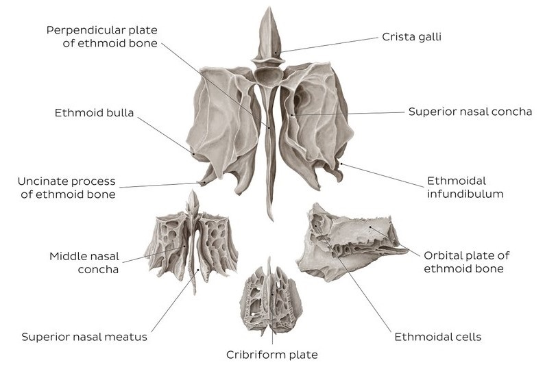
The ethmoid bone is a 3D square bone situated at the focal point of the skull between the eyes. It helps structure the dividers of the eye attachment, or orbital pit, as well as the roof, sides, and inside of the nasal cavity.
Exceptionally light and wipe-like on the surface, the ethmoid bone is one of the most intricate bones of the face.

Table of Contents
Ethmoid Bone Function
Since the ethmoid bone is in the face, its role is to assist in a variety of regular exercises. The cribriform plate has strainer-like openings that permit the olfactory nerves to exist in your nose so you can smell things and enable you to taste them.
Sinus pits in the ethmoidal maze assist with serving numerous significant capacities, including:
- Fluid creates within the body to trap allergens and foreign particles that are harmful when taken in through the nose.
- Vocal tone
- Decreasing the heaviness of the head
- The nasal conchae that the ethmoid structures permit air to flow and become humidified as it goes from your nose on the way into your lungs. The bodily fluid that delivers in the sinus cavities lines this piece of your nose, which likewise fills in as a safeguard component by catching any particles that might cause ailment or different responses.
Ethmoid Bone Structure
The ethmoid bone is a front cranial bone between the eyes. It adds to the average mass of the circle, the nasal pit, and the nasal septum. The ethmoid has three sections: cribriform plate, ethmoidal maze, and opposite plate. The cribriform plate shapes the top of the nasal cavity and adds to the development of the foremost cranial fossa. The ethmoidal maze comprises an enormous mass on one or the other side of the opposite plate. The opposite plate frames the predominant 66% of the nasal septum. Between the orbital plate and the nasal conchae are the ethmoidal sinuses or ethmoidal air cells, which are a variable number of little cavities in the sidelong mass of the ethmoid.
Enunciations
The ethmoid explains with thirteen bones:
There are two bones in the neurocranium – the frontal-facing bone and the sphenoid (at the sphenoidal body and the sphenoidal conchae).
Eleven bones of the viscerocranium – two nasal bones, two maxillae, two lacrimals, two palatines, two mediocre nasal conchae, and the vomer.
Development
The ethmoid hardens in the ligament of the nasal case by three focuses: one for the opposite plate, and one for every maze.
It is during the fourth and fifth long periods of embryonic life that mazes first develop, with ossific granules appearing in the lamina papyracea and spreading into the conchæ.
Upon entering the world, the bone comprises the two mazes, which are small and poorly created. During the third year after birth, the opposite plate and crista Galli start to solidify from a solitary place. Further, they join to the mazes about the start of the subsequent year.
Cribriform plates harden halfway from the opposite plate and partially from the mazes.
The development of the ethmoidal cells starts during fetal life.
Embryology
The ethmoid bones grow principally through endochondral hardening. The ethmoid bone begins to frame at around 25 to 28 weeks of growth. Ethmoid sinuses are available upon entering the world. Dissimilar to all of the other sinuses, the ethmoid bone creates from the cartilaginous olfactory container or paleosinus while the other paranasal sinuses stretch out from the ethmoid into the membranous bone through epithelial diverticula expansions.
Certain stages are essential for the advancement of the paranasal sinus, which incorporates essential and optional pneumatization designs that call for differential development of the cartilaginous olfactory case. These stages create diverticular pockets that extend inside the limits of the container, which manufacture elaborate intracapsular aviation route spaces.
Auxiliary pneumatization designs happen inside the paranasal break, where the diverticula leave the limits of the nasal container to become extracapsular. They are left consuming space in nearby designs, making the paranasal break become a paranasal sinus.
Even though the size of the ethmoid bone becomes controlled, the extents of the neosinus (which incorporates the front-facing, maxillary, and sphenoidal) can change fundamentally because paranasal breaks are not uniform. The pace of advancement or separation in the extracapsular diverticula relocation isn’t steady, and when the extracapsular diverticula become extramural. They extend beyond the limits of the ethmoid bone and pneumatic close-by designs to produce different waste pathways.
Clinical Significance
The ethmoid bone is significant in the underlying arrangement of the skull and plays a defensive part in the human body. The cribriform plate permits the olfactory nerve to go through from the nasal chamber into the skull. Pneumatized ethmoid air cells guarantee primary soundness, opposing breaks inside the average mass of the circle. This also safeguards the eyeball during injury. Orbital breaks are related to numerous tasteful deformities, for example, enophthalmos, diplopia, facial lopsidedness, hypertelorism, and an oppressed world.
Enophthalmos, which is the regressive and descending uprooting of the globe into the hard circle, happens all the more normally when the lamina papyracea cracks. When there is herniation of the orbital substance in the ethmoid.
NOE
Since the naso-orbital-ethmoid (NOE) complex is a sensitive anatomic design, clinical assessment and radiographic examination are basic to survey the place of the globe compared with the orbital edge, eyelid edge, and other facial designs. NOE wounds might start from hard impacts during quarrels or engine vehicle mishaps, and pupillary errors, retrobulbar hematoma. The optic nerve harm precludes in such wounds
Ethmoid breaks might bring about postponed complexities even a very long time after injury; nonetheless, such cases are somewhat uncommon. These intricacies incorporate meningitis, a repeat of cerebrospinal liquid rhinorrhea, and deferred cerebrospinal liquid rhinorrhea beginning. A past report found that patients with these difficulties frequently have enormous ethmoid bone imperfections. Moreover, they frequently have ordinary radiological evaluations after injury. With the fruitful careful fix, these patients were well after follow-up.
Patients with irritation of the ethmoid bone might have osteitis of the ethmoid bone and constant rhinosinusitis. Constant rhinosinusitis happens when the nasal and paranasal sinus mucosa becomes aroused for longer than 90 days without a complete side effect goal. The practical endoscopic sinus medical procedure (FESS) is the therapy for this condition. The motivation behind FESS is to open up the sinus air cells and sinus Ostia with an endoscope.
CT sweeps of patients with constant rhinosinusitis have displayed to have areas of expanded bone thickness and unpredictable thickening of the sinus dividers. This could bring about a repeat of the sickness after careful therapy and act as wellsprings of ongoing inflammation.
Previous concentrates on recommended patients going through endoscopic sinus medical procedures for persistent rhinosinusitis may have osteitis of the ethmoid bone. The pervasiveness could also be essential as high as 64%. Meningitis or a sore connected with osteomyelitis of the ethmoid bone may likewise bring about anosmia or taste aggravations.
FAQs
These consist of the matched parietal and fleeting bones, in addition to the unpaired front-facing, occipital, sphenoid, and ethmoid bones.
According to antiquated texts and customs, mind expulsion was the first phase of the treatment. A snared instrument is used to break the ethmoid bone, which isolates the cerebrum from the nasal depression. A similar device was used to remove the cerebrum and related tissue.
